Site Menu:
| This is an archived Horseadvice.com Discussion. The parent article and menus are available on the navigation menu below: |
| HorseAdvice.com » Diseases of Horses » Skin Diseases, Wounds, and Swellings » Swellings / Localized Infection / Abscesses » Diagnosing and Assessing Swellings in Horses » |
| Discussion on Swollen Mass In Front of Udder...Cellulitis or Something Else? | |
| Author | Message |
| Member: dr3ssag3 |
Posted on Friday, Aug 31, 2007 - 5:37 pm: Greetings, Everyone.After spending a small fortune on I-RAP (which appears to be successful), my dear Contessa has invented yet something else to have me worried about. A few months ago, she developed what I mistakenly thought was a fat pocket in front of her right udder. I thought little of it as she has a tendency of developing such pockets whenever she's laid up. However, my vet noticed that it was a hard mass...too hard for it to be a fat pocket. He did some bloodwork, which showed only that if anything her blood work was a bit low. We decided to see if it went away with exercise. It did not. Two weeks later, my other vet (from the same practice) was out doing her teeth and decided to do an ultrasound. Whatever he saw inspired him to withdraw whatever fluid was in it (about 6ccs) and send it off for testing. He also managed to extract a small piece of tissue for biopsy. The test results are apparently vague enough to make him want more tests...a larger area to ultrasound, rectal exam, etc...a five-hour appointment I have to haul in for next week. I asked for copies of the reports so that I could post them in case anyone else has seen this sort of thing before. Sadly, they were faxed so I only have them in TIF format, so I'll be manually typing the condensed version...as this message is becoming rather lengthy (sorry). Here goes: From the tissue sample: "MICROSCOPIC: Sections from the submitted specimen of tissue contain fibrin and adipose tissue with scattered eosinophils and degenerate mononuculear cells, small well-differentiated lymphocytes, occasional macrophages and hemorrhage. DIAGNOSIS: Mixed cellulitis with fibrinous necrosis, eosinophilia and hemorrhage, ventral abdomin, lateral to right mammary gland. COMMENT: Sections from this specimen contain mixed inflammatory infiltrates with eosinophilia fibrin and hemorrhage. Culture is indicated based on evaulation of this sample. These sections lack evidence of signficante cellular atypia or neoplasia. Additonal biopsies may be warranted if there is clinical suspicion of underlying disease processes." And for the Fluid Sample: "MICROSCOPIC: Refractometer protein <2.5gm/dL. Nucleated cellularity is low to moderate, comprised primarily of small mature lymphocytes with occasional eosinohils, lymphoblasts, rare macrophages. INTERPRETATION: Lymphocytic Inflammation. COMMENTS: Results indicate either a site of lymphocytic inflammation or sampling from a reactive and edematous lymph node, or potentially a dialted lymphatic vessel. There is no sign of malignancy, suppuration, or infection. Histopathologic evaluation may provide more information. Further cultures will not be set up based on cytology; histopathology results are pending." I've been doing all the research I can but unfortunately I've only got an MA in English...I don't do vet speak very well as science always made me dizzy. All I know is that Contessa appears to be fine in every regard...she's eating, drinking, has not lost or gained weight, and is her general irritable mare-ish self. The only thing I have noticed is that this summer her coat has been "ruddy"...usually she's extremely shiny as she gets 1/2 cup corn oil daily. I'm not sure whether to be worried or not, so of course I'm erring on the side of worry and driving myself and my saintly husband mad. Any and all insight will be most appreciated. Thanks, Dawn |
| Member: mrose |
Posted on Saturday, Sep 1, 2007 - 8:46 pm: Hi Dawn! IMHO I wouldn't be too worried...easy for me to say because it's not my mare. As long as there's no sign of cancerous growth, and it doesn't sound like there is, I wouldn't be too worried. Of course, I'm not vet. Dr. O may say something that might make me want to eat my words of encouragement, but I hope not. |
| Member: dr3ssag3 |
Posted on Monday, Sep 3, 2007 - 3:47 am: Thanks, Sara. Every encouraging word helps. I guess i always get spooked when they call for "more tests." My vets tend to err on the side of extreme precaution...something I appreciate but can, in times like these, find taxing on the nerves.Oh and i noticed in my original post that I accidentally put "blood work" in where "white blood cell count" should have been. Dawn |
| Moderator: DrO |
Posted on Monday, Sep 3, 2007 - 10:22 am: Dawn it reads like an old site of trauma that has not completely resolved. Is the area painful or hot as these may indications of something more serious? What is your veterinarian's concern?DrO |
| Member: paul303 |
Posted on Tuesday, Sep 4, 2007 - 1:48 am: I own a mare with a mass that resembles your physical description, Dawn. It developed, I believe, in 2002 - at least, that is when I first noticed it. We sent the mare to New Bolton for a biopsy - it was unsuccessful - the young vet was fearful and couldn't administer a local. She DID suggest that we have her checked for Cushings....we were a bit miffed at that time and basically just blew her off. Our vet did a biopsy and sent it out and our result was sort of like yours ( as I remember ), non-definitive. I've lost track of the results ( I tried to find it for another thread a couple of years ago ), they were copied and sent out for a few opinions but we never pinned it down. The size of the mass changed in the beginning, yet it was never soft or movable. It's remained the same for a few years now. It's never given her any trouble, nor is it sensitive or painful.After a couple of years, we DID have her checked for Cushings - she was exhibiting symptoms. She was diagnosed with Cushings and has been on Pergolide for 3 years. I've always wondered what made the young vet in New Bolton suggest a Cushings test ( when the mare was there for a biopsy of a lump ). At that time, my mare was about 22, bright, rotund, with a glossy perfect coat and no thrush or founder type foot problems. Yet, within a year or two, she started to show Cushings symptoms. Now, did that young woman have ESP, or was she somehow making a connection with Cushings and the tumor like mass? I had myself in such a snit that day that I wouldn't take the time to talk to her.....I just might have learned something. |
| Member: dr3ssag3 |
Posted on Thursday, Sep 6, 2007 - 1:15 pm: I don't think that it's particularly painful and there's definitely no sign of heat. Since she's back to work, and since the vet extracted fluid it seems to have reduced slightly in size.I believe my vet just wants to be sure that there isn't something internal going on that would cause this to manifest. They plan on doing a 10cm(?) ultrasound as the ultrasound they had with them at the farm was only 6 cm (?)... I'm not sure what the exact measurements are... They also want to do a rectal examination. We're going to be hauling her in today so I'll hopefully know more tonight. Dr. O, is it possible that this is Cushing's? She shows no signs indicative of Cushing's and I'd thought that tumors/growths associated with Cushing's were limited to pituitary(sp) glands. Contessa's issue most likey deals with the mammary gland, correct? Thanks, Dawn |
| Moderator: DrO |
Posted on Friday, Sep 7, 2007 - 8:00 am: I don't associate skin or body tumors with Cushings but without examination cannot say what the association with the mammary gland might be.DrO |
| Member: dr3ssag3 |
Posted on Friday, Sep 7, 2007 - 2:41 pm: The vet visit yesterday was insightful, but there's still no name for what she's got.The vet wanted to rule out any tumors etc by doing the ultrasound and rectal exam. Nothing irregular showed up. Apparently, the fungal cultures came back positive, although they still don't know what exact type of fungus is there. They'll be checking the cultures again in a week's time. Meanwhile, another blood draw was performed along with a belly tap. The fluid in the belly tap looked good, although she thought she saw something floating in it. I'm curious about the fungus thing...if anyone's seen anything like it. It appears to be localized in the mass itself, but I'm confused as to what might have caused her to get it, and how other horses that have been exposed to her haven't come down with anything strange. Obviously we'll know more once we get a definitive assessment of the cultures. Contessa also showed problems with allergies for the first time this year. The mass appeared over the winter, but when the vet was out for IRAP she had these horrible itchy welts from bug bites found exclusively in the region around her udders and the mass. There was pitting edema on the mass itself. The vet put her on HyDrOxyzine and the welts went away and have not come back. I'm wondering if the two are linked and if so, how? Could allergy issues be to blame? Dawn |
| Moderator: DrO |
Posted on Saturday, Sep 8, 2007 - 8:34 am: Usually deep fungal infections of the skin come from wounds Dawn but a light culture might be a contaminant particularly since no fungi showed up in the fluid exam. This could be a severe reaction to a bug bite and if so I guess you could call it an "allergic reaction".DrO |
| Member: paul303 |
Posted on Sunday, Sep 9, 2007 - 1:38 am: Mmmm boy, the mare I mentioned above, had a miserable cullicoides allergy, and used to rub her ventral midline raw in the season. When she was 18 years old, we changed locations and the "sweet itch" vanished. The lump in front of her udder developed not too long after the move. She spent some time on corticosteroids to try to control her allergy - this was prior to her move. Interesting. |
| Member: dr3ssag3 |
Posted on Tuesday, Sep 11, 2007 - 3:59 am: A phone call from my vet today reveals another answer that begets further questions.The fluid from Contessa's belly tap was normal. However, her white blood cell count has gone a bit lower from July (down to 4.3). Although she appears healthy apart from the mass, she's showing signs of being slightly anemic, but it's not an iron deficiency, more like "anemia of chronic inflammation/disease"... (I'm researching this next.) My vet is still waiting on the most current results for the fungal culture (to be taken Thursday), and pending those results we might test for Cushing's just to rule it out, if nothing else. Outwardly, you see a mare in perfect health (aside from the mass). Although her coat seemed dingier than last year, it exhibits none of the Cushing's symtoms. She is anything but "depressed" and/or listless, and (knock on wood), we've never had any issues with laminitis. The only abnormal thing this year is the allergies...but what I've seen does not appear to be anything like what one sees in "Sweet itch" cases. She had a few isolated welts that immediately responded to hyDrOxyzine and have not reappeared. Dawn |
| Moderator: DrO |
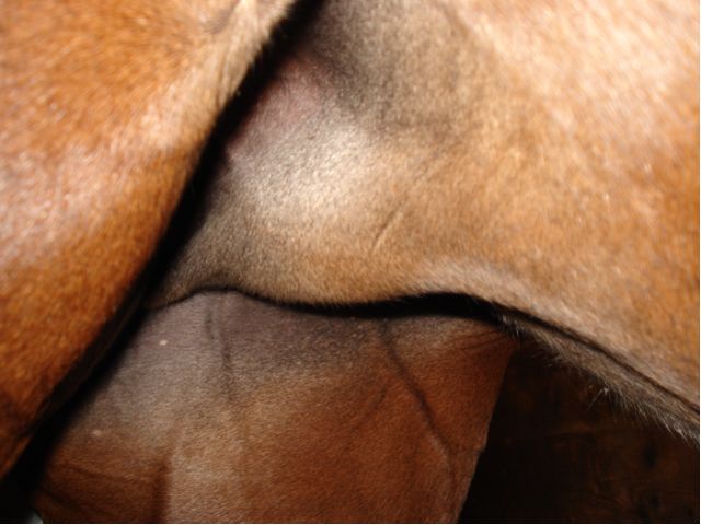
Posted on Tuesday, Sep 11, 2007 - 7:02 am: Dawn, can we see a image of the swelling?DrO |
| Member: dr3ssag3 |
Posted on Wednesday, Sep 12, 2007 - 11:55 pm: I've attached a couple of images taken today. The mass is approximately 3" in length and about 1 1/2" at the widest point. It seems to have gone down in size since she's been back to work.   The left side has a small fat pocket that does not exhibit the same firmness as the right side. 
|
| Moderator: DrO |
Posted on Thursday, Sep 13, 2007 - 8:55 am: Location is a bit further back than I had pictured earlier and considering the rich lymphatics at this point dilated lymphatics possibly secondary to trauma and/or occlusion of the drainage would be at the top of my rule out list and unlikely to be of any consequence. If I had a client very worried about the uncertainty of the diagnosis I would consider ultrasound to better define the swelling and the surrounding vasculature and rectal to explore the inguinal canals.I would consider DMSO (+/-dexamethasone) and light massage as therapy. DrO |
| Member: dr3ssag3 |
Posted on Monday, Sep 17, 2007 - 3:39 am: I'm going to run this by my vet to see if it's an avenue she'd like to explore. I noticed on Thursday it seemed a bit larger and there were more welt/bug bites on it. She'd also begun rubbing her tail again.After speaking to the vet, we decided to up her hyDrOxyzine to 500mg (? - 10 tablets) twice daily instead of just once. Tonight when I looked at it it seemed ever so slightly smaller and definitely cooler to the touch. |
Horseadvice.com
is The Horseman's Advisor
Helping Thousands of Equestrians, Farriers, and Veterinarians Every Day
All rights reserved, © 1997 -
is The Horseman's Advisor
Helping Thousands of Equestrians, Farriers, and Veterinarians Every Day
All rights reserved, © 1997 -
