Site Menu:
| This is an archived Horseadvice.com Discussion. The parent article and menus are available on the navigation menu below: |
| HorseAdvice.com » Diseases of Horses » Lameness » Joint, Bone, Ligament Diseases » Arthritis and DJD: An Overview » |
| Discussion on Xrays finally... | |
| Author | Message |
| Member: Sunny66 |
Posted on Tuesday, Mar 22, 2005 - 10:23 am: 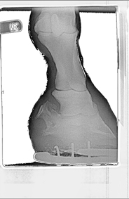
|
| Member: Sunny66 |
Posted on Tuesday, Mar 22, 2005 - 10:29 am: 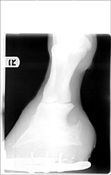 Vet said he wouldn't go above training level with him...if that. He said he's looking at the big picture and I can make my own decisions. He said the more I ride him the faster he'll go downhill. He gave him 5 years before complete retirement. Do I retire him now and get another horse....or ride him for awhile, then get another horse? He said I'm doing all the right things. I've had poly shoes put on his front feet to ease concussion, I'm having his back shoes pulled Wednesday. Xrays show the farrier is doing well...only mm's off at places. I can upload those too if you like. What is your take on this Dr. O, have you seen worse but have a successful riding career? |
| Moderator: DrO |
Posted on Tuesday, Mar 22, 2005 - 11:21 pm: Aileen the pictures are too small for me to make any detail out on. Could they me made larger? Start with the originals and just reduce them to a width of 500 pixels, maintaining the aspect. Then reduce the image to just below 64K. We are traveling right now but I will look at them this weekend.DrO |
| Member: Sunny66 |
Posted on Wednesday, Mar 23, 2005 - 10:39 am: I'm having difficulty, but try this one. I could also email them to you, that way you'll get the full xray. Let me know if that's ok. |
| Member: Sunny66 |
Posted on Wednesday, Mar 23, 2005 - 10:41 am: Here's another one in case this works.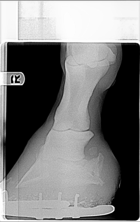
|
| Member: Sunny66 |
Posted on Wednesday, Mar 23, 2005 - 10:44 am: 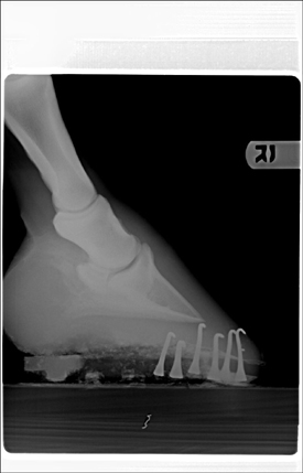
|
| Member: Sunny66 |
Posted on Wednesday, Mar 23, 2005 - 10:52 am: So sorry, it looks like I double posted one. Please delete one if you like...These were taken 3.21.05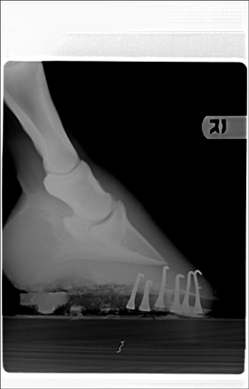
|
| Moderator: DrO |
Posted on Friday, Mar 25, 2005 - 10:20 am: I can see he has remarkable calcification of the lateral cartilages (probably not significant) and the 10:41 image appears to have something going on around the coffin joint, but I cannot make out any detail.These images were originally 8 by 10 and for the detail we need, for the type judement you want, it is going to require a full size original on a screen, only then can I compare these lesions with those I have seen in the past. Let's try to get this on firmer ground. What lesions did your veterinarian diagnose from the above radiographs that made him make the above statement? DrO |
| Member: Sunny66 |
Posted on Friday, Mar 25, 2005 - 11:18 am: Thanks Dr. O,I believe he was diagnosing the coffin joint. Yes the sidebone is significant, and I have another vet looking at these who has seen his xrays before to see if there has been much change to the sidebone as well their view on the ringbone. I'll be happy to email you full size xrays. I understand there may be a consult fee. Where should I email them? And how many do you want me to send? |
| Moderator: DrO |
Posted on Friday, Mar 25, 2005 - 5:21 pm: Aileen what is their significance from a lameness standpoint? With the caveat that there may be lesions not visible in this format, I have seen many radiographs of horses with side bones as bad or worse that were sound.I will email you with information on charges and where to send the radiographs for such a consultation over the weekend. DrO |
| Member: Sunny66 |
Posted on Friday, Mar 25, 2005 - 7:36 pm: The sidebones didn't seem to be bothering him, but maybe after his rodeos they did...how would I determine that? The vet seemed fairly confident that his issue is ringbone.The sidebones seem to have grown a bit to me...I don't remember them being THAT high, but yes, they were high. I'll look for your email. Thanks again Dr. O. |
| Moderator: DrO |
Posted on Saturday, Mar 26, 2005 - 9:11 am: The side bones can be palpated. Pain on deep palpation that blocks out with a unilateral ab. sesamoid block on the side of the suspect sidebone would be a good start.DrO |
| Member: Sunny66 |
Posted on Saturday, Mar 26, 2005 - 11:32 pm: Would I be able to palpate the sidebones? If so, how exactly would I do that?On another note...he is not quite right. He's eating his grain, but not very into his timothy hay, but it is cleaned up by morning. This has been going on all last week. We had a tremendous storm last week. This is usually his prime colic/impaction time. He doesn't seem to be dehydrated, he's drinking well, and with his watery stools I'm assuming I don't need to worry about impaction...but who knows. His pulse is 40 -- which I'm gathering is ok since he hurts -- has gut sounds, more on the right side than the left. He has watery stools...not quite diarea (sp?) but he swishes his tail after he poops, so it must sting. Of course my thermometer isn't working and it's too late now to run to the store to get another. He was peeing, but only a trickle came out this morning; however, later on he peed a decent one. I haven't seen him do a racehorse pee in a long time. The vet said last week he had a dirty sheath. I have a call into him to come out this week - hopefully sooner than later - and clean it, I didn't want him tranquilized last week because he was already in a huff...I have never seen my horse so angry and we were just taking xrays. My question to you is -- what do I give him if it's not his sheath causing him discomfort? He's on probiotics. I've heard to give him bran, but I wanted to check with you first, it seems to me that the bran would increase the watery stools. Or is it the fiber he needs? I'm in the process of switching him from omalene 100 to straight oats. I'm in the third week of transition. He's getting 1.5 cups oats and .5 cups omalene. Could this be why and I shouldn't even be worrying? I just want the poor guy comfortable and I don't know what to do. |
| Member: Sunny66 |
Posted on Sunday, Mar 27, 2005 - 10:40 am: I didn't give him probiotics last night, and this morning his stools were well-formed. I know, I know...I'm micromanaging. |
| Member: Sunny66 |
Posted on Monday, Mar 28, 2005 - 10:23 pm: Hi Dr. O,I haven't received an email stating where to send the xrays yet. |
| Moderator: DrO |
Posted on Tuesday, Mar 29, 2005 - 10:42 am: I am sorry Aileen, I should have just made this simple email it to horseadvice@horseadvice.com.DrO |
| Member: Sunny66 |
Posted on Tuesday, Mar 29, 2005 - 10:53 am: No problem. I've emailed you.I did speak with the vet last night and he said as long as he's sound he can gallop around the pasture...but not until then. He said "this is an injury and it needs to heal" ... I'm confused STILL about this...I thought ringbone does not ever heal, but just gets worse? Am I making any sense? I very much appreciate you taking the time to look these over. |
| Moderator: DrO |
Posted on Tuesday, Mar 29, 2005 - 11:22 am: You appear to be making sense but I don't understand the vets thought about degenerative joint disease of the coffin bone bone healing: this is chronic arthritis. Are you sure you are on the same wavelength with your vet?Looking at your radiographs only raises more questions. Looking at rf2, which is an oblique of the foot, at first glance looks like changes consistant with coffin jt arthritis but...we have to remember that the lateral cartilages are ossified. For those follwing this discussion it is the 10:41 am radiograph above. I cannot be sure we are not seeing the overlying lateral cartilage, making it appear that the joint has developed remarkable osterphytes. In fact I find this a likely explanation but would require further radiography to prove or disprove. I don't see evidence of DJD of the coffin joint evident on the lateral (straight from the side) or ap (the one taken directly from in front of the foot) and the other obliques are too underexposed to read this aspect of the study. Though we do not see it in the other views, often the first place to see these changes are in the oblique shots. I recommend the vet take a series of obliques that slowly work around the foot to differentiate these 2 possiblilities. Yes you can palpate the side bone. Look on the ap radiograph and it will show you exactly where to feel them. DrO |
| Member: Sunny66 |
Posted on Tuesday, Mar 29, 2005 - 11:36 am: I think I have more xrays of the right front...I'll send those to you tonight and see if you can see from that. Otherwise, the vet is coming out again next week, and I'll ask for more.Are you saying he may not have articular ringbone? What is an osterphyte? Sorry I'm at work and can't look it up. |
| Member: Sunny66 |
Posted on Tuesday, Mar 29, 2005 - 2:14 pm: So it could be basically a bone spur? If this is the case, does it change his riding career outlook? For better or worse? I guess this would be one of my key questions.Another key question would be, can he PLEASE go out to pasture and at least be a horse? |
| Member: Sunny66 |
Posted on Tuesday, Mar 29, 2005 - 11:37 pm: I've sent you two more xrays Dr. O. Let me know if you can see these any better...if not, I'll ask the vet to do the obliques next week.Thank you! |
| Moderator: DrO |
Posted on Thursday, Mar 31, 2005 - 7:44 am: Right, he may not I cannot tell if the messed up looking joint is actually the joint or the overlying side bone superimposed on the joint. This may be discernable on the originals. Yes whether your horse has DJD of the coffin joint might have a significant effect on the prognosis. Since the diagnosis is not clear from these radiographs Aileen I cannot guess proper treatment.DrO |
| Member: Sunny66 |
Posted on Thursday, Mar 31, 2005 - 10:19 am: I completely understand Dr. O, and I also want you to know that I would not necessarily take your diagnosis to heart, but would greatly value your opinion. I also have another vet -- his old lameness vet -- looking at the xrays and am waiting to hear what he says as well.If it is indeed ringbone, which I would hope he wouldn't have said so if it were questionable since it's such a dire prognosis - and suggested and done the injection in his coffin joint on February 23 -- would you have recommended stall rest and handwalking? I've been doing this since Feb. 23. Now that the rain has stopped, FINALLY, I'm sure his lameness will be minimal and he may even be sound, he's turning much better now; however, I don't feel qualified to say if he's sound now. He is getting a little punchy, and I don't blame him one bit. I've started giving him access to his 50 x 70 turnout in the mornings only -- knowing he's very quiet in the mornings. I've done this just last weekend and he just walked and walked and walked...and he was better because of it. I do, however, want to know how you feel of his prognosis of riding training level only. I have heard people say that they have bought a horse with ringbone and competed successfully for 10 years and then retired the horse at 20. I know it depends on the horse, since my guy is so sensitive I am in no way assuming this would happen for me, but I would like to know if continuing to ride him would cause him to not be with me as long as he would be if I didn't ride him at all anymore. Another take on this would be that since this is arthritis, riding would be better for him than just pasture...this is what I was told about his hocks, so I'm wondering if this would also apply to the articular ringbone. I do understand that while you are incredibly knowledgeable, you cannot predict the future, but your input would be greatly appreciated. |
| Member: Sunny66 |
Posted on Saturday, Apr 2, 2005 - 3:28 pm: Hi Dr. O, I've been reading again!How exactly would I determine if my horse has DJD of the coffin joint or just coffin joint lameness? Another round of radiographs? How long would I have to wait to determine this? If you look at the xrays I sent to you ...rf1 is from Feb. 23 and rf2 is from March 21. I may be just hoping tooooo much, but doesn't it look better in rf 2?? ...and couldn't it be better because he is not getting so much trimmed off? Having finally figured out that my horse has been trimmed WAY too short for the past year and a half, could this contribute to this condition? My farrier will from now on take off next to nothing to keep his foot well aligned. You can actually see the difference between the hoof growth in these two xrays. RF1 there is a definate slant, in RF2 it is much more even. |
| Moderator: DrO |
Posted on Saturday, Apr 2, 2005 - 6:16 pm: DJD of the coffin joint is said to be present when lameness is localized to the coffin joint and radiographic changes show arthritis is present. Without good radiographic evidence isolating it to the coffin joint exactly is not a simple matter but explained at, Equine Diseases » Lameness » Diseases of the Hoof » Overview of Diagnosis and Diseases of the Foot.It is not another "round of radiographs" you need you just need necessarily to find out if that the irregular radiopague areas overlying the joint in rf2 are actual changes in the joint or due to the overlying side bone. If that takes a few more obliques from that side or your vet interpreting the radiographs he has I don't know. DrO |
| Member: Sunny66 |
Posted on Saturday, Apr 2, 2005 - 6:57 pm: Thanks Dr. O,I'll read up on it tonight and hopefully have my vet out this week. My horse feels good and is moving much better at the walk -- really walking out now -- with only a very slight headbob to the right at the trot while turning - but he seemed sound and happy on the straightaway (not on purpose mind you - he took it upon himself). He's also turning tightly MUCH better than he has in over a month. Crossing my fingers... |
| Member: Sunny66 |
Posted on Wednesday, Apr 6, 2005 - 10:21 am: Vet's coming today for another round of xrays to see if what I saw on the farrier series of xrays is correct...that it is getting better... the more hoof he has, the more aligned his hoof is, the less dramatic coffin joint issue.Please wish us luck! |
| Member: Sunny66 |
Posted on Wednesday, Apr 6, 2005 - 6:22 pm: I've sent you three more xrays Dr. O.Vet said: That it is ringbone, not the collateral cartiledge. That he is sound on the straight, sound on a circle to the left but lame (1 out of 10) to the right at a trot. I'm still confused....he said that the ringbone is quieting down...better by 10% to 15%. Again, how could it get better if it's ringbone?? Vet said still no turnout or if I do ...not to tell him. I have been turning him out in his 50 x 70 pen that attaches to his stall run when it's warm and toasty and there isn't a storm brewing, but only in the mornings since he's sleepy -- and he said he's getting better. Farrier was here as well. I showed her the xrays the last time she was here. Showed her how the coffin bone was then in line with the sole. She shod my horse after the vet took the xrays. She took off almost 1/2 inch of toe and barely any heel today while I was talking to the vet. I looked at the xrays taken today, Dr. O. In them his sole was perfectly even with his coffin bone. I am at my wit's end. Please tell me that this isn't detrimental to my horse's way of going due to his injury. I wish I had the money to fly you out to California so you could see my horse in the flesh. I'm so frustrated. |
| Member: Dres |
Posted on Wednesday, Apr 6, 2005 - 6:39 pm: Aileen, are you any where near UCD... they have a great farrier that works off x rays...On the first day God created horses, on the second day he painted them with SPOTS.. |
| Member: Sunny66 |
Posted on Wednesday, Apr 6, 2005 - 7:28 pm: Thanks Ann,I'm about 1.5 hours from UCD...can you email me the name? adalen@m-w-h.com It's always good for me to know of a good farrier - even though I'd probably have to trailer out. I just talked to my friend and told her what I stated about the farrier and she said basically that the farrier had to take off hoof in order for him to be aligned still in 7 weeks. If she just let him go untrimmed his angles would be totally off by the time he was shod again. As long as she took off evenly all the way around he should be just as aligned...However, the farrier told me she took off toe and left his heels. His feet look small again and I think part of his progress is because they were allowed to grow. I know I'm not a farrier, but if were asking these questions more in the past, maybe I'd still have a sound horse. |
| Moderator: DrO |
Posted on Thursday, Apr 7, 2005 - 10:02 am: Assuming we are talking about the same thing, I disagree with your veterinarian. If you look closely at the side bone in the first (lateral) radiograph there is a little notch at the top. In the second (oblique) shot the area that looks like severe DJD changes of the coffin joint also has a notch at the top: they look like the same notch to me.I don't think the relationship of the coffin bone to most of the sole is bad, but agree with the farrier, the toe needed chopping off in front. Just off the front, I would not thinning the sole uner at the toe. DrO |
| Member: Sunny66 |
Posted on Thursday, Apr 7, 2005 - 10:34 am: Ok, so based on your view, assuming you're leaning toward a bone spur. What would your treatment be? Stall rest, turnout?THANK YOU...I feel SO much better getting your opinion on the way he is shod...MUCH appreciate you Dr. O!!! Don't forget to tell me how much and where to send it. |
| Moderator: DrO |
Posted on Friday, Apr 8, 2005 - 9:41 am: No not a bone spur, in one of the obliques what looked like severe osteoarthritis of the coffin joint appears in these later images to be artifactual, and instead the superimposed calcified cartilages. Given that I am loking at images in a low resolution environment, I don't see evidence of coffin joint arthritis.DrO |
| Member: Sunny66 |
Posted on Friday, Apr 8, 2005 - 10:09 am: Thanks Dr. O,I've received another viewpoint similar to yours from his lameness vet (they will not travel to me, I have to trailer him out - which is the only reason why I used a different vet as he has toys on his truck): "I am not convinced of the Dx of ringbone. I am assuming that you mean "low ringbone," which involves the DIP joint (coffin joint). The PIP joint looks good. I see that he has significant sidebone, which will not cause lameness, but is often an indication of imbalanced feet or shoeing. The radiographs that you sent, IMO, are not diagnostic of the DIP joint. There is considerable overlap of the sidebone on the obliques, and the DV view is too "light:" to adequately assess the coffin joint. If he is talking about some apparent resorption of cortical bone seen on the lateral, I would have to compare that to the physical exam, ie, results of flexion tests, local blocks, and articular blocks. We would need more information to assess this case. In any case, there is not significant radiographic evidence of ringbone shown on the attachments that you sent to me." Based on both of you agreeing...do you feel stall rest is still necessary given the latest information of him only being slightly off to the right on a circle at the trot? I do have yet another vet (his overall vet) coming out on the 21st for spring stuff and a lameness exam as well. And depending upon what the 21st brings, once my horse is acting normally again I will trailer him to my lameness vet. |
| Member: Sunny66 |
Posted on Friday, Apr 8, 2005 - 11:11 pm: Hi Dr. O,Could you point me in the direction to find out what exactly this means? apparent resorption of cortical bone seen on the lateral Thank you! |
| Moderator: DrO |
Posted on Sunday, Apr 10, 2005 - 8:15 am: Aileen you really are going to have to ask him what he means because he seems to be referring to something the first vet might be thinking.Resorption of cortical bone means there has been loss of calcium or bone mass and not something I find in the images of the radiographs I have but would best be assesed on the originals. DrO |
| Member: Sunny66 |
Posted on Sunday, Apr 10, 2005 - 9:20 am: I'll ask. Can you tell me what exactly does this mean as far as my horse is concerned? Is this something that can be fixed? Can you speculate on the prognosis if this is the case?Thank you Dr. O... |
| Member: Sunny66 |
Posted on Sunday, Apr 10, 2005 - 11:07 am: Ok, I found some information, whether this pertains to my horse...I don't know.Is the bone in his hoof basically remodeling itself? If so, is movement better or is stall rest better for him? |
| Member: Sunny66 |
Posted on Sunday, Apr 10, 2005 - 2:09 pm: I talked to the vet today, he said that the resorbtion of the cortical bone is most likely due to the fact he hasn't been fully weightbearing on the foot. Does that make sense?On a great note: He trotted sound both ways today! I let him trot today since he felt like it. Head down to the ground shaking it...not once did his head come up and I surely wasn't going to ask him to raise it... Of course I'll still have the vet out and look him over before I get too excited. Is it true that SOME horses show ouchiness in their feet when it is actually their tummies hurting? He's had corn oil now for 4 days -- I had heard that may help coat the lining of the stomach and decrease acidity -- and papaya for two days -- another thing I heard that may decrease acidity. It is highly possible I know that this lameness has just run it's course - or that he'll be lame in a couple of days or even tomorrow - so I'm still going to have the vets out. |
| Moderator: DrO |
Posted on Sunday, Apr 10, 2005 - 6:03 pm: There is no doubt that with decrease use that bones remodel, but trying to evaluate bone density from a set of field radiographs is, in my opinion, is a bit or a reach. I don't think this is a significant problem with lameness on even rehab unless you plan or racing this horse.Delighted to hear about the horse feeling better. I have never seen a lameness / colic differential diagnosis problem. DrO |
| Member: Sunny66 |
Posted on Sunday, Apr 10, 2005 - 9:04 pm: Please accept my heartfelt appreciation and gratitude for putting up with me through all this.I'll update after the 21st to see if the other vet can see the ringbone in the xrays. If he can't, I'm going to forever wonder what it really was. I wish the vet would have ultrasounded when I asked. |
is The Horseman's Advisor
Helping Thousands of Equestrians, Farriers, and Veterinarians Every Day
All rights reserved, © 1997 -
