Site Menu:
| This is an archived Horseadvice.com Discussion. The parent article and menus are available on the navigation menu below: |
| HorseAdvice.com » Diseases of Horses » Lameness » The Interpretation of Radiographs » |
| Discussion on Need Help Interpreting Right Front Radiograph | |
| Author | Message |
| Member: divamare |
Posted on Friday, Jan 27, 2012 - 2:44 pm: The pastern joint doesn't appear to match the angle of the coffin joint or the fetlock joint...does it?What does this suggest? What else in these X-Rays would suggest 'trouble'? Mare is three and now in corrective shoes. Medial and Lateral balance was terrible. She is in a show barn and not worked a great deal and has little turn out. Thank you everyone. 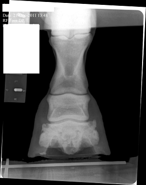 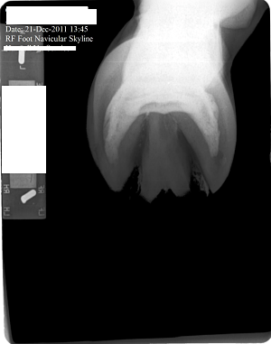 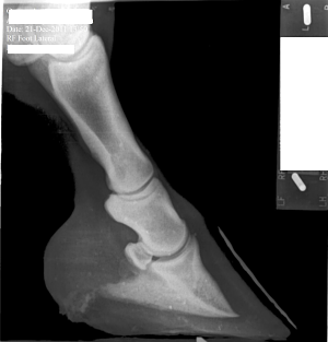
|
| Member: divamare |
Posted on Saturday, Jan 28, 2012 - 9:52 pm: Anybody home?
|
| Member: canderso |
Posted on Sunday, Jan 29, 2012 - 8:15 am: Hi Vicki,I think we are all waiting for Dr O... I could tell you what I see and what I think it means, but I am not a vet and you would be foolish to listen to me! Sit tight - it is worth the wait. Cheryl |
| Member: divamare |
Posted on Sunday, Jan 29, 2012 - 7:53 pm: Thanks Cheryl. It was late Friday when I posted, and I figured everyone would be off having a week end. Anyway, for what it's worth, I see the beginning possibly of side bone on the lateral side. I see the pastern joint angle does not match the angles of the fetlock and coffin joint. I cannot tell where the coffin joint is in relation to the coronary band. She has long under run heels and long toes. I don't know if the depth/shape of the collateral grooves tells me anything. The ski tip on the front of the coffin bone bothers me especially on a horse only 3. And is there a small bit or rotation down as well? I don't understand in the X-Ray why her sole appears to be bearing the weight and the hoof walls are off the ground. Is this really how she was prior to 'corrective' shoeing or is this X-Ray poorly taken or am I just poor at interpreting it. Or has her hoof mechanism sunk into the hoof capsule some? I do not have vet remarks for these X-Rays. |
| Member: divamare |
Posted on Sunday, Jan 29, 2012 - 8:10 pm: 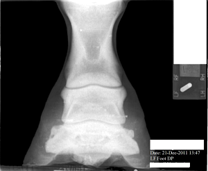 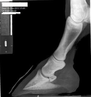
|
| Member: divamare |
Posted on Sunday, Jan 29, 2012 - 8:16 pm: left front The first X-Ray cut off the fetlock joint. This was not an editing error.The coffin joint looks uneven to me. Bigger space on the medial side. What does that mean? Nothing or something. I can't tell if this mare is built like a stack of playing cards or not(remember stacking cards like the wobbly Leaning tower of Pisa?). And how come the left lateral view the hoof wall is on the block but the right lateral view the hoof wall is off the block...is this an operator error or wonky foot? |
| Member: dres |
Posted on Monday, Jan 30, 2012 - 10:29 am: i have no clue how to read rads ... what makes u believe u see the beginnings of side bone?On the first day God created horses, on the second day he painted them with spots.. |
| Member: divamare |
Posted on Monday, Jan 30, 2012 - 12:03 pm: Ann, look at the left lateral radiograph. The small hook at the top, lateral side of the coffin bone. It sticks up a wee bit higher than the other side and is perhaps? moving a bit past the beginning of the coffin joint. As I understand it sidebone isn't always related to a lameness issue, and many older horses have it. However, this is a 3 yr old. I "think" it is a minor issue... |
| Member: dres |
Posted on Monday, Jan 30, 2012 - 10:23 pm: Thank you Vicki... |
| Moderator: DrO |
Posted on Tuesday, Jan 31, 2012 - 8:12 am: Hello Vicki,In general I avoid interpreting radiographs on the internet as the quality of the images here are not high enough to do a good job and the amount of time required to look at all aspects of the radiograph. I do invite members to put up radiographs and I will comment on any diagnosis made by the veterinarian who took the radiographs. That said I do think the RF foot in the front appears to have an overly long toe and corresponding toe flare and underrun heel. I don't see these issues in the L fore. The problem with assessing the conformational issues your bring forth is that technique and how the radiographs are taken can greatly effect the relationship between the bones and create false impressions. For instance all a horse has to do is stand a little forward over the foot and the normal pastern will not be aligned. The conformation issues you raise are best assessed looking that the horse while standing square and moving in a straight line than by a static set of radiographs. DrO |
| Member: divamare |
Posted on Thursday, Feb 2, 2012 - 12:21 am: Thank you very much. |
Horseadvice.com
is The Horseman's Advisor
Helping Thousands of Equestrians, Farriers, and Veterinarians Every Day
All rights reserved, © 1997 -
is The Horseman's Advisor
Helping Thousands of Equestrians, Farriers, and Veterinarians Every Day
All rights reserved, © 1997 -
