Site Menu:
| This is an archived Horseadvice.com Discussion. The parent article and menus are available on the navigation menu below: |
| HorseAdvice.com » Diseases of Horses » Eye Diseases » Topics on Eye Diseases Not Covered Above » |
| Discussion on Marks | |
| Author | Message |
| Member: Contilli |
Posted on Friday, Jan 20, 2006 - 9:22 pm: My vet just left and is unsure what is in the lenses of my 4 year old geldings eyes. He has two marks in the 'lens' of his left eye. The retina looks good. He is not sensitive to light, blinks when stimulated and seems perfectly fine - other than becoming head shy. His right eye has a similiar mark at the 9:00 mark. His left eye (in the photo below) has marks at the 12 & 6 locations. The marks are greyish in color. The circle at the bottom of they eye is from my flashlight.... Do you have any idea what this may be? The retina looks good.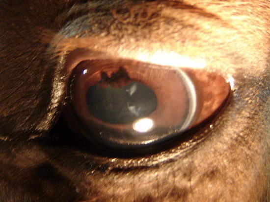
|
| Member: Mrose |
Posted on Saturday, Jan 21, 2006 - 10:23 am: I know nothing about the marks, but am really impressed with your photographic skills. |
| Member: Paul303 |
Posted on Sunday, Jan 22, 2006 - 2:35 am: That was my very thought, Sara. An extraordinary picture, Denise.I had a gelding that developed a distinct sword-shaped white slash suddenly. It didn't look like your's. It was about 1/2 inch long, and 1/4 to 1/8 inch wide, with soft edges and started from the lower outside corner extending across the iris to about the pupil. He displayed no symptoms. Vet said to watch carefully - most likely corneal abrasion. I watched it for what seemed like forever, then gradually got used to it being a part of him. Eventually, it went away. I doubt it was the same thing, however. |
| Member: Angel77 |
Posted on Sunday, Jan 22, 2006 - 2:47 am: Dear Denise,I am also impressed by your photography. I care for a 29 yr old mare with the same type of-I do not know what it is. It doesn't seem to bother her. It is in both eyes. Located dead center over the cornea(I think that is the correct term?). Please keep us posted. I would like to know the diagnosis. Nice dialation of the eye. Good Luck to you and your boy. WTG |
| Member: Contilli |
Posted on Sunday, Jan 22, 2006 - 9:18 am: Thanks for the complements. It was a challenge to hold him, the flashlight and the camera about 1/2 inch away from his eye! But I got a couple of good shots.My vet still has not come up with anything. I asked him to draw blood for Lepto titers which he will do on Monday. I've also emailed the photos to Marion duPont Scott Equine Hospital and New Bolton. No answers yet. Max is my Dressage Prospect which I bred and have waited 4 years to ride. He is quite spectacular and, quite frankly, I am freaking out at this point! I would have to say these marks DO impair his vision. He is becoming head-shy and sometimes when I am walking towards him with a flake of hay, he backs up and arches his neck like he's not sure what I've got... His eyes were checked in August and they were clear of any marks. I know Dr. O is busy, but where is he :-) I'd like him to respond to these photos....... Thanks everyone. Denise www.BryantFarm.com |
| Member: Contilli |
Posted on Sunday, Jan 22, 2006 - 10:05 am: This is his left eye and the mark is slight and around the 9:00....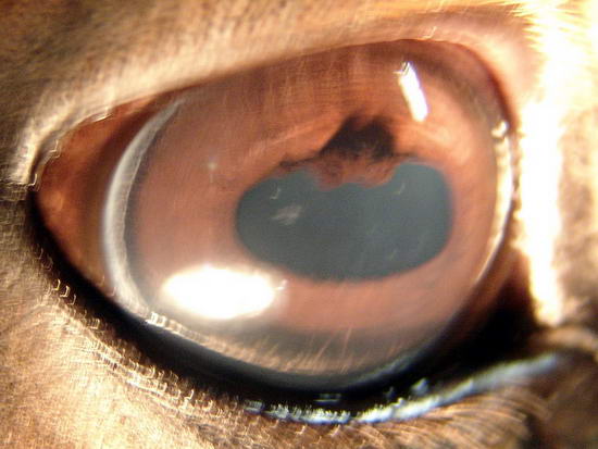
|
| Moderator: DrO |
Posted on Sunday, Jan 22, 2006 - 5:57 pm: Hello Denise,These are cataracts and the prognosis depends on their appearance when viewed with an ophthalmascope and the level in the lens where they occur. This may require a slit lamp examination. As explanation of how to classify these by appearance / location and the associated prognosis see, Equine Diseases » Eye Diseases » Cataracts in Horses. I agree with those above that the photos are super however they will may appear different in an ophthalmascope when back lit by the reflected light of the retinae. Let us know what your vet says on the exam. By the way what color and breed is this horse. DrO |
| Member: Contilli |
Posted on Sunday, Jan 22, 2006 - 6:13 pm: Hi Dr. O:Thanks for your input. My Vet did an eye exam with his handheld eye/light opthalmascope - the instrument you mentioned. The marks are deep in the eye and in the lens. Max is a 4 year old Warmblood Dark Chestnut. What do I do next? What machines/tools/probes do I ask for? Thank you, Denise www.BryantFarm.com |
| Member: Lilly |
Posted on Sunday, Jan 22, 2006 - 9:18 pm: Denise,Have you made an appointment with a horse optometrist yet? My barn manager mentioned that there is a good one in Richmond, VA. I can get the name for you if you are interested. Ann |
| Moderator: DrO |
Posted on Monday, Jan 23, 2006 - 7:23 am: Hmmmm...deep in the eye or lens does not help me: are they cortical or nuclear (again see article for definitions) is what is important along with the appearance in the scope.No matter what is wrong a very viable option is to keep an eye on them and see if they enlarge you can have that fancy eye exam then. There are no early treatments for cataracts (that are not secondary to inflammation and yours do not appear to be) that I am aware of. I think it is more likely these are not progressive and do not significantly effect sight than they are a problem. If you are not a wait and see type person make a appointment with a veterinary ophthalmologist who has a slit lamp he can carry into the field. DrO |
| Member: Vickiann |
Posted on Monday, Jan 23, 2006 - 3:50 pm: Is your Warmblood imported from Germany? My daughter has a ten year old Grand Prix jumper from there (Holsteiner) who has marks in his right eye. The Vet who did the check prior to purchase said they were nothing to worry about -- just old scars. Unfortunately there were (a few months later) some flare ups of inflammation, treated with banamine and antibiotic ointment. Because the condition began to come back when treatment was stopped, the stable manager where my daughter keeps him took the horse to the University of Florida where he has seen a couple of specialists. The diagnosis was "superficial Keratitis." Some special ointment was prescribed. Any new flare ups are to be taken seriously and treated. The doctors feel this can be managed in a way that will control the situation without damaging the eye permanently. An interesting remark the Dr. made was that he only sees this condition in imported warmbloods (I THINK German Warmbloods). If I have posted this in the wrong place I apologize, but when I saw "chestnut warmblood," felt I should respond. |
| Member: Contilli |
Posted on Monday, Jan 23, 2006 - 5:30 pm: Hi Vicki-No, he is not from Germany, I bred him myself. His mother and siblings have clear eyes. Interesting about your horse. Glad it is treatable. I think the lens may be too deep for eye ointment but antibiotics may due. Also, a friend asked me if my builders were welding recently. Because she feels they appear to be welding marks. And, as a matter of fact they were. I went to the barn to find them repairing a piece of equipment. I thought to myself "could this hurt my horse whom seems to be staring at the welding process?" I thought - I am being too paranoid! So, I left it alone. My neighbor wonders if him staring at the welding has caused this damage? I do have an appointment on Wednesday at 10am with an ophthalmologist. I will post her findings asap. Thanks for all your support! Denise |
| Member: Vickiann |
Posted on Monday, Jan 23, 2006 - 7:08 pm: WOW -- That is amazing. I found out this UF Vet, previously mentioned, was talking about Warmbloods coming from Europe in general, not just Germany and the marks are strange scars, supposedly caused by previously active periods of the "superficial keratitis. I hope you will have a good outcome with your horse and think it is great you are taking him to an ophthalmologist. Good luck to you -- early and correct treatments are so important with eye problems. |
| Moderator: DrO |
Posted on Monday, Jan 23, 2006 - 8:14 pm: Denise is correct there is a difference in changes in the cornea (keratitis) and changes in the lens and the two problems not comparable.DrO |
| Member: Contilli |
Posted on Tuesday, Jan 24, 2006 - 12:41 pm: This is the response from Leesburg Hospital (they saw the photos not the horse):Hi Denise, Thanks for the photos - they are pretty impressive since more often than not taking photos of the horses eye is a nightmare with the light bouncing straight back at you. Anyway I have got all sorts of people to look at them here at the hospital and the general consensus is that we feel a second opinion is needed. The spots look to be on the back of the lens and our number one concern would be that he has a posterior uveitis and the spots are fibrin tags. If that is the case then the condition will progress unless addressed. Obviously feel free to give me a call if you have any other questions (7037716800) but we would be more than happy to see him if you wanted to bring him to us. Possibly see you soon, All the best, Dr. Rachel Atherton |
| Moderator: DrO |
Posted on Wednesday, Jan 25, 2006 - 7:03 am: Thanks for the update Denise,I would not have said that determination could be made looking at these photos, at least I cannot. But if an ophthalmologist thinks this is likely, best to follow their advice and be sure to get a copy of the final report and pass it along to us. DrO |
| Member: Contilli |
Posted on Wednesday, Jan 25, 2006 - 7:57 am: Inclement weather has caused me to miss my appointment today. But, my twin sister works for a vet in California who looked at the photos and felt that they are lesions from a head injury or hitting his head in the past. He, the vet, has one in his eye from a head injury. He says that his floats around.... Interesting.... I will be taking Max to Leesburg Hospital tomorrow, they have an indoor facility but not an ophthalmologist. They do have slit lamp and plenty of vets around to 'rule out' stuff. Will post more :-) |
| Moderator: DrO |
Posted on Thursday, Jan 26, 2006 - 7:03 am: Good luck Denise,DrO |
| Member: Contilli |
Posted on Thursday, Jan 26, 2006 - 3:10 pm: A very sad diagnosis for a 4 year old!Diagnosis from Marion duPont Scott Equine Medical Center 1/26/6 Dr. Desrochers & Dr. Atherton: Degenerative changes to the lens bilaterally/early cataract development Freiheitsfest 'Max' presented to the Equine Medical Center today for evaluation of opacities visible within the lens of both eyes. At presentation he was bright and alert with seemingly normal vision. Fluorescein and Rose Bengal stains were negative bilaterally. Ophthalmic examination highlighted the presence of opacities within the lens of both eyes with four being present in the left eye and two smaller ones in the right eye. Ultrasound of both eyes confirmed the lesions to be located within the lens and in both eyes mild floating hyperechoic areas were visible within the viteous possible consistent with prior or current inflammation. In the left eye the anterior capsule of the lens showed more severe degenerative scelerotic change. Thanks everyone! This is a photo of Max's left eye via Ultrasound. 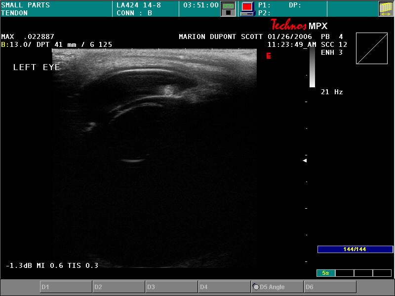
|
| Member: Mrose |
Posted on Thursday, Jan 26, 2006 - 3:31 pm: Am I reading this right, that inflammation caused this? What if anything can be done to slow down the progression? Can cataract surgery be done on horses as it is on humans?I feel so sorry for you, Denise. I know you are very attached to this guy. Did the doctors give any recommendations or hope at all? |
| Member: Contilli |
Posted on Thursday, Jan 26, 2006 - 3:54 pm: Hi Sara:Most commonly Uveitis is the culprit for changes in the lens. BUT, Max has never had ANY symptoms of Uveitis. I bred and raised Max and believe me, if he was wincing from the light, had runny or itchy eyes or looked as if in pain I would have known it. Max is a total wimp to pain and I'm with him every day, 3 - 5 times a day. So, no, inflammation is not the culprit. I am totally convinced that he has not and does not have Uveitis. Our Lepto Itiers will be back from Cornell this week. I was surprised that they can't tell..... if there is inflammation... They are not saying what has caused this. Foals can be born with Cataracts but have a 50-60% vision recovery rate when the lens is removed before one year of age. Thank you for your sympathy! You are correct - I am attached to him. I kept him out of all my foals because he had the most potential! Denise www.BryantFarm.com |
| Member: Dres |
Posted on Thursday, Jan 26, 2006 - 5:09 pm: Denise, has the vet given you any further instructions to help him.. wearing a fly mask in sunshine or windy days.. ?? Am so sorry to hear your news.. I know how you feel, I have a gelding that was an FEI prospect, he is now turning 7 and been lame off and mostly on now for three of those years... he is my pasture pal and love him dearly, but heart broken...On the first day God created horses, on the second day he painted them with spots... |
| Member: Mrose |
Posted on Thursday, Jan 26, 2006 - 7:22 pm: I don't know why it is, but it often seems like things happen only to "the best." Our mare that blew out her knee was qualified for Nationals when she did it, and she's a beautiful mare that we bred and raised. We still love her, but like yours Ann & Denise, she had such great prospects! Just makes you want to either cry or scream! (or both!) |
| Moderator: DrO |
Posted on Friday, Jan 27, 2006 - 8:08 am: Denise, take some heart in that I have seen such dire pronouncements in other horses with no history of inflammatory events, that never had any problems or at least the problems manageable. For instance the pronouncement of degenerative sclerotic change suggests findings that would be difficult to distinguish on ultrasound from a static defect that formed during intrauterine development. Keep us appraised of developments.DrO |
| Member: Contilli |
Posted on Friday, Feb 3, 2006 - 5:55 pm: Update -Just received the results back from Cornell Uni. Negative for Lepto! So, can he have inflammation without Uveitis? Ultimately, Uveitis is caused by being exposed to Lepto right? Could there be bilateral posterior Uveitis due to inflammation without being exposed to Lepto? I am now confused. Thanks, Denise |
| Moderator: DrO |
Posted on Saturday, Feb 4, 2006 - 8:27 am: Hello Denise,The relationship between Lepto and RU is explained at Equine Diseases » Eye Diseases » Anterior Uveitis, Recurrent Uveitis, Periodic Opthalmia, and Moonblindness. The literature occasionally describes chronic inflammatory changes where no acute flares have been observed but I have not seen one and I find it hard to believe that the first sign is a cataract deep in the lens. DrO |
is The Horseman's Advisor
Helping Thousands of Equestrians, Farriers, and Veterinarians Every Day
All rights reserved, © 1997 -
