- This topic has 22 replies, 2 voices, and was last updated 1 year ago by
clauee.
- AuthorPosts
- May 24, 2024 at 9:49 am #21778
clauee
MemberHi Dr O
I have a 3 yo lusitano filly who has been gradually losing weight and muscle in topline for one year now, yet her belly expands. In short, she looks like a very old broodmare. I first noticed the subtle changes when she was about 20 months old.She has never been sick, no fever or depression or lack of appetite, and has normal energy for her age. She lives in a small group of horses on 16 acres and gets along very well with her herdmates.
Initially we revised all the feed (which didn’t require any change, the filly eats good quantity and quality for her age), and did some bloodwork including vitaminE and selenium, tested genetic PSSM and wormed twice despite negative worm counts. All was completely normal but she kept losing muscle and weight very slowly and gradually, while her belly kept expanding. I even thought she could be in foal at one point!
Then this spring the vet noticed a grade 2 heart murmur. It is diastolic, left, mitral. It is difficult to hear so it could have been easily unnoticed before..
I asked for further testing as I thought there could be a relationship between the muscle loss and the finding of heart murmur. Also I noticed she has not grown at all in the past 8 months. So we proceeded to exercise tolerance tests and checked the muscles enzymes (normal) and complete neurological exam (normal).
We finally found some clues at the abdominal ultrasound. It appears that the liver is enormous… so big the vet doesn’t understand how the filly can eat normally as it takes all the space for the colon. Abnormal tissues extend beyond her bellybutton! Also the last blood test revealed elevated liver enzymes which matches the ultrasound findings. However the numbers are not very high for now.
We proceeded to a liver biopsy (echo guided) yesterday and should have results within two weeks. At first glance the vet mentioned the abnormal tissues don’t look like liver, but rather like fat (white and brittle). However a “fat liver” has been excluded from the possibilities as the filly is very lean.
The vet said at this point possibilities for treatment are practically null. It appears to be a chronic pathology and something very rare for sure.
Any thoughts of what this could be? I have read elsewhere about a disease called “hepatoblastoma” which seems to correlate best with the symptoms and findings for now.
I don’t know how the help my filly…For now she shows no sign of pain whatsoever but we are expecting that she will show signs of abdominal pain in a near future if this keeps expanding.
- May 25, 2024 at 11:32 am #21781
 Robert Oglesby DVMKeymaster
Hello clauee,
We should wait until the biopsy results come back for the symptoms you describe are very nonspecific. Liver enlargement, hepatomegaly, could be acute inflammation, neoplasia, toxicity, secondary to heart disease, congenital, and others. The swollen abdomen is most likely excess peritoneal fluid. Is the CBD and bilirubin normal? Characterizing the fluid as either inflammatory, exudate, or transudate may help substantially.I would like to address some of the abnormalities you discuss. First liver enzyme elevation is not always an indication of liver failure and large changes can occur with relatively mild insults to the liver. A mild heart murmur is also a common finding and may not indicate serious illness. So these changes may or may not be related to the main problem. The serious finding is weight loss in the face of good nutrition and deworming program. Let’s see if the biopsy suggests a diagnosis and if not consider further diagnostic testing like a peritoneal tap to check what is in the excess peritoneal fluid which may lead us to liver function tests (bile acids, bilirubin), checking for heart disease, and perhaps elsewhere.
DrO - May 25, 2024 at 8:11 pm #21783Member
Hi DrO
Thank you for your response.The blood results are all normal (bilirubin) except for elevated AST, GGT and GLDH but these numbers aren’t very high nor suggestive of hepatic failure. The vet said that these results for a horse with no clinical signs would require another control in two weeks and not be too worried about it.
She also did not see any fluid in the abdomen at the ultrasound. Only an enlarged liver and liver tissues extending very far in the belly. She even mentioned it would be very difficult to collect fluid for a test.
I will give an update once we have the biopsy results - May 30, 2024 at 1:21 pm #21786Member
Hello dr O
The vet called and said the biopsy result is… Fat. Only fat.
The team at the university hospital don’t understand how this is possible. The filly has no fat over the ribs, how can she get some so deep inside the abdomen? This also means that the liver can’t be distinguished from these fatty tissues on ultrasound.
We did another bloodwork and all is normal except again the same three enzymes a bit high (ast, ggt and GLDH). The first two came down a little compared to 3 weeks ago, while the GLDH doubled. However the result remain “low” at 43 so it’s still not really worrisome but we need to find the cause behind this!
The team will proceed to another biopsy and try to find some liver. They will also do transrectal palpation. They are consulting colleagues as this seems very “interesting” medically and for sure extremely rare. - June 1, 2024 at 12:48 pm #21787Robert Oglesby DVMKeymaster
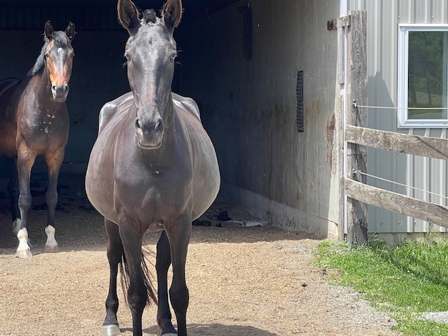
clauee, was there any explanation of the type of fat found? Excessive visceral fatty tissue in the face of a declining body condition places endocrine involvement like hyperadrenocorticism, excessive steroid administration, or possibly thyroid disorders on the rule-out list. If she were older (much older) pituitary adenoma would be top of the list. Could you post an image of your horse? One from the side and one from behind. Also, an outline of what and how much she is fed and supplements would be helpful along with the recent history of changes.
DrO - June 2, 2024 at 7:06 am #21788Member
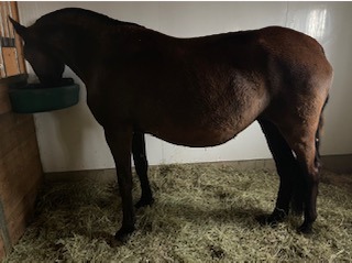
Hello dr O. Here are pictures while she still had winter coat.
She eats free choice grass hay 24-7 (between 9-11% protein and HNCS +/- 10%). A stall test revealed that her intake on 24h is approximately 22-23 pounds and that makes sense with what I see from her in the field and the amount of poop I muck every day. Her poop is normal.
She gets a daily supplement of 200g beet pulp mixed with alfalfa + Madbarn Omneity complete supplement. I add some commercial feed usually Purina for young horses (200g – 400g depending on season), salt, vegetable oil in winter (1/2 cup) and fresh grounded flaxseed during coat changes.
There is a free choice vitamin/mineral block available at all times along with salt blocks.In summer she is on pasture.
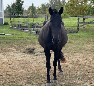
She has no problem losing her winter coat unlike PPID horses. I added a photo of last summer after pasture season. At that stage the muscle loss had been going on for 6 months and wasn’t as pronounced as now. Her bloodwork were completely normal including selenium and vitamin E.
One of the university professors has a student working on a microbiome analysis for horses. They asked to collect from my horse to compare to their dysbiose index. I hope this will give more clues.
I try to post pictures but the size is too big. I’ll see how I can figure this out
-
This reply was modified 2 years ago by
-
This reply was modified 2 years ago by
- June 2, 2024 at 7:24 am #21790Member
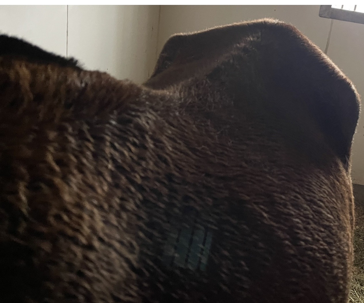
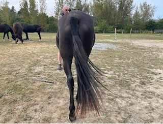
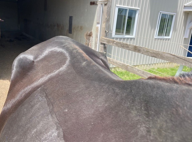
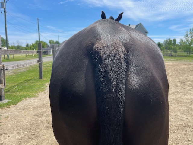
The pictures you see are from last September and the first two ones are recent (April)
- June 2, 2024 at 7:54 am #21795Member
History of changes:
– April 2023: first notice of muscle loss along topline and mostly croup. The filly is 21 months old.
– September 2023: no improvement despite Sumer pastures and careful review of feed. Bloodwork including vitamin e /sélénium normal. Neurological exam normal. Genetic testing for PSSM negative. The vet notices a slight heart murmur. The filly has an expanding belly making her look pregnant.
April 2024: i notice the topline being more atrophied including the back and more weight in the belly. We do another complete neurological exam (normal) and muscle tests after effort (normal) and exercise tolerance test to monitor the heart murmur. It is graded as left/mitral/diastolic grade 2. Doesn’t get worse after exercise. Ultrasound reveals abnormal tissues in the abdomen and bloodwork comes back with slight elevated GGT, AST and GLDH. The vitamin e is slightly low. Sélénium normal. We do a liver biopsy.
– May 2024: results of liver biopsy reveal fat and second blood test reveal an elevation of GLDH (43) and lower AST/GGT which are still slightly above normal. Further exams will take place (second biopsy, rectal palpation and microbiome analysis). Bloodwork on other horses of the herd are all normal (no elevated liver enzymes).
- June 2, 2024 at 11:51 am #21796Robert Oglesby DVMKeymaster
Wow, the loss of condition is remarkable. Besides a biopsy of the liver, pursuing the possibility of heart disease and endocrinopathies would appear to be a rational course. A muscle biopsy could help in finding the cause of the wasting.
We have a weight loss topic that contains many articles on weight loss in horses: https://horseadvice.com/horse-equine/diseases/colic-diarrhea-gi-tract/weight-loss-in-horses/. Still, I agree none of the odd weight loss diseases fit exactly: Chronic Grass Sickness, Proliferative Enteropathy, Chronic Ionophore Poisoning or Chronic Selenium Toxicosis come to mind.
As to what else you might be doing consider, continuing with free choice hay and mineralized salt block but switching out the assortment of supplemental items with a good quality complete pelleted horse feed with 16% protein. The amount of pelleted feed fed should be increased until weight improvement is seen. I am assuming all the other horses get the same water and hay.
DrO - June 2, 2024 at 1:27 pm #21797Member
Hi DrO
Here are pictures today.
Yes all the horses are fed the same water (municipal and human consumption quality) and hay, but they are currently on fresh pasture. We are in Canada so the pasture season is mid May-mid September. All the other horses have good bloodwork and normal weight, one horse of the herd is fat but is also IR.-
This reply was modified 2 years ago by
Attachments:
-
This reply was modified 2 years ago by
- June 17, 2024 at 4:19 pm #21827Member
Hi Dr O
We got the results and the filly is not IR nor Cushings
The vets don’t know what is going on
- June 18, 2024 at 10:10 am #21828Robert Oglesby DVMKeymaster
When equestrians say “Cushing’s” that is a bit of a misnomer. Equine Cushings is not truly Cushing’s Disease but instead pituitary dysfunction. True Cushings is hyperadrenocortisim which PPID mimics. Did they test for pituitary dysfunction (common in older horses) or adrenal dysfunction (rare but not unknown)? What about thyroid disease? I would still like to see a peritoneal fluid analysis.
Your horse is losing condition unexplainably. Your vets need to list possible causes and rule them out one by one. If they cannot find out what is happening a referral to an equine internest is the next step. Have you made the changes in the diet recommended above? How is that going?
DrO - June 18, 2024 at 3:59 pm #21829Member
Hi Dr.O
When I say « the vets » I mean the university hospital vets (which is what I think you refer to as equine internist… sorry my english is not so good!). There is one team on the road for clients in a close range. It’s been a while that my local vet referred this case to them!The test was ACTH but the filly has no symptoms whatsoever and we were not surprised by the negative result. The insulin test is baseline insulin and was 3,6.
At this point the equine specialist says that we could do eventually peritoneal fluid analysis or a laparoscopy to explore the fatty tissues, or a muscle biopsy. However they think these tests won’t reveal much in terms of knowing « why » she is like this or give any possibility of treatment. They also mentioned the paracenthesis would be difficult because there is so much fat everywhere the needle wouldn’t be long enough. So for now they said to keep up with the diet and try to do a little more exercice but they don’t think it will help much.
Since they have no clue what is going on, and especially that the filly is doing very well despite her weird shape (she has good energy, interacts normally, no pain anywhere on the body, good coat and hooves etc) it doesn’t match any known disease. As for the thyroid test I asked and they said the result interprétation is not reliable? - June 18, 2024 at 7:42 pm #21830Member
Hi dr O
I checked the correct titles of the vet in charge of my mare and it’s « internal medicine service clinician » and « equine hospital medical chief » at University of Montreal veterinary practice. In my area it’s the highest level of medical equine services we can get. She assessed my mare several times and did the liver biopsy too.
She says this is a very particular case and not anything seen before. - June 18, 2024 at 7:57 pm #21831Member
As for the diet, we’ve been increasing protein as much as possible in the last year (with no results and despite the fact the diet was considered above the needs for protein content).
I’m waiting for a new diet plan from the nutrition specialist based on all the recent lab results. - June 20, 2024 at 7:44 am #21834Robert Oglesby DVMKeymaster
clauee, I am having trouble with the logic of “we don’t know what is going on” yet “don’t feel further diagnostics helpful”. I understand that we all have limited resources, and once they are played out, we have done all we can. I am not sure where they (you) are at.
Concerning thyroid testing, simple thyroxin levels are not helpful but T3 suppression tests can be. The best information we have on your situation will be found at https://horseadvice.com/horse-equine/diseases/colic-diarrhea-gi-tract/weight-loss-in-horses/ and I would start with the Overview article as it provides a step by step approach to the problem.
DrO - June 20, 2024 at 11:13 am #21835Member
Hi Dr O
Thank you for your response. I’ll review the article.The combination of clinical signs and test results give an overall portrait that the vets don’t understand.
The ultrasound is the most concerning as there is a thickness of visceral fat everywhere which makes other tests like paracenthesis and liver biopsy difficult or non-diagnostic. They also don’t see any liquid on ultrasound. They don’t believe any other available test will explain « why » these fatty tissues/masses are there and most importantly how to reverse that.
I am extremely confused myself as to what to do best for this mare…
I’m open to further testing if the tests can reveal a cause for which there is a treatment. So it depends what we are looking for…
Otherwise I don’t want to stress the filly with invasive procedures and trips to the hospital away from her herd. My priority is her quality of life.
So this is not a financial limit, I’m just trying to find the balance between her quality of life and the possibility of a diagnosis with positive outcome.
- June 21, 2024 at 5:00 pm #21841Robert Oglesby DVMKeymaster
clauee, review the article on Malabsorption in Horses, https://horseadvice.com/horse-equine/diseases/colic-diarrhea-gi-tract/weight-loss-in-horses/malabsorption-in-horses/. I was updating it with case study review and your horse came to mind.
- June 22, 2024 at 7:21 pm #21845Member
Thank you. I will read it!
- August 2, 2024 at 11:49 am #21896Member
Update:
A new vet from Guelph equine hospital came to assess the filly.Ultrasound: same diagnosis. The liver cannot be seen on right side as the visceral fat is too thick, even to the maximum possible setting of depth view (I believe 30cm if I understood well). A biopsy is there has very little chance of finding liver tissues.
The fatty tissues extend everywhere in the abdomen and lots are towards the omentum. On the right side the liver can be seen in a very small spot and does not have a normal texture at all. A biopsy there would be too risky and not recommended.
Heart murmur: could no longer be heard. However the heart beats are unequal in intensity – sometimes loud and sometimes very weak.
Rectal palpation: normal.
Blood results: steady. Liver enzymes slightly elevated but everything else normal.
Paracenthesis was tempted but unsuccessful to collect fluid.
Other test options (that were decided not to proceed):
– laparoscopy : invasive and would not give treatment options
– muscle biopsy : there are no clinical signs of muscle disease other than the loss of topline (no tying up, trembling or trouble getting up/down). Already has been done genetic testing for muscles diseases, and blood work after effort is normal. The lifestyle and diet with liquid vitamin E supplementation is already done to cover for muscle diseases that we can manage. Any other findings with a biopsy would not give a solution nor an explanation to the accumulation of visceral fat.
-glucose absorption test: the filly has no clinical sign of malabsorption other than the loss of topline. Blood work is all normal and she is alert, good hair/hooves/energy etc. It doesn’t fit with the accumulation of visceral fat which seems to be the main issue.
For now the filly is still pain free and normal in every aspect of her life (appetite, energy, curiosity etc).
- August 2, 2024 at 12:13 pm #21897Member
I found this article which seems to fit with most of the test results we have so far, but in this case the horse was obese:
https://journals.sagepub.com/doi/pdf/10.1177/104063879500700435
- August 2, 2024 at 3:29 pm #21898Robert Oglesby DVMKeymaster
Hmmm, a rapidly growing fat cell neoplasia, that histologically appears as normal fat cells, that is invading the abdomen. It is causing limited demonstrable organ damage while creating a negative energy metabolism. Pretty far out there, but as you say it fits the exam and lab findings so far. I wonder why the a difference in metabolism effects between the horse in the report and your horse? Abdominal lipomas are common in horses but tend to be limited in size and often pedunculated. The “stem” often gets rapped around a loop of the bowel creating a strangulating colic. I have lost an aged horse that way. However, the horse in the report coliced from complications of the damage to the intestinal wall. Perhaps your neoplasia has more affected the ability of the bowel to absorb nutrients than the weakening of the wall.
DrO - March 6, 2025 at 4:10 pm #22180Member
Hi drO
A last update on this case. The mare was euthanized because the feces became smaller and smaller (although normal in consistency and frequency, with no signs of colic) and there was subtle ventral edema for the past weeks. Other than that she was in good shape, still running around with the other horses and showing no sign of pain and good appetite. Bloodwork was normal except for the slightly elevated liver enzymes that remained steady in the past year.First findings of autopsy is a huge (extremely huge) fatty tumor originating from the omemtum/mesentery. We gave the body to the university for further studies.
- AuthorPosts
- You must be logged in to reply to this topic.