Diseases of the Splint Bones in Horses
by Robert N. Oglesby DVM
Introduction
Introduction
»
Diagnosis
»
Splints
»
Distal Splint Fractures
»
Proximal Splint Fractures
»
More Info & Discussions
The splint bones are two small long bones that lie along each side of the cannon bone in both the front and rear legs. Those in the front are called metacarpals (Mc), and in the rear metatarsals (Mt). These are fairly easily palpated just in front of the edges of the suspensory ligament. They are vestiges of the 2nd and 4th digits of the early horses millions of years ago. The inside splint bone (2nd Mc and Mt) is usually longer than the outside (4th Mc / Mt) and has a more extensive articulation at the knee or hock. Toward the bottom the splints taper smaller until some 1 cm from the end when they flare out into the 'bulb or button' which is easily palpated in most horses. In young animals, a fibrous interosseous ligament unites the splint bones with the cannon which calcifies progressively with age.
These bones are fairly easily fractured and are also susceptible to excessive concussion from work in the young horse. This article discusses diagnosis, treatment, and prognosis of diseases of the splint bones.
Diagnosis
Introduction
»
Diagnosis
»
Splints
»
Distal Splint Fractures
»
Proximal Splint Fractures
»
More Info & Discussions
|
|
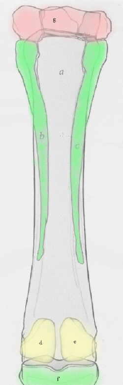
Image of the Cannon (a) and Splint Bones (green b & c) from the back of the cannon.
|
Diseases of the splint bones are usually seen as local swelling which may or may not be accompanied by heat and pain over the splint bone. These swellings can have two presentations:
-
Swelling not associated with lameness. Splints that present at this stage have minimal soft tissue swelling around them and are not remarkably painful on palpation. However if they are not rested till cool they can become painful.
-
Swelling associated with a forelimb lameness. Usually there is soft swelling over a more firm swelling on the splint bone. The area is painful on palpation. Occasionally the lameness precedes the swelling by several days so a splint should be considered with any occult lameness.
Radiographs are important when lameness accompanies a new splint. The radiograph will show an area of periosteal swelling, new calcium deposition, and possible may reveal a fracture if the splint is from trauma. Splints are radiographed using oblique views of the cannon with a relatively low setting. Technique that illuminates the cannon bone will overexpose the splint, so they must be radiographed separately.
Chronic, old, or cold splints are seldom a source of lameness and are usually noted as a cosmetic blemish. Occasionally the bony mass will be felt to impinge on the suspensory ligament. However, if a lameness exists, the splint should not be incriminated unless other causes have been eliminated by careful use of regional anaesthesia.
Splints
Introduction
»
Diagnosis
»
Splints
»
Distal Splint Fractures
»
Proximal Splint Fractures
»
More Info & Discussions
To read more on this topic become a member of
Horseadvice.com! Your membership gets you instant access to this and over 600 equine articles on our site. Other benefits of your membership include participation in our discussion boards and access to our one button PubMed search tool for each topic.
Horseadvice.com educates you to be a more knowledgeable horse owner which leads to healthier horses and save you money, we guarantee it. Come Join Us!