Overview of Anatomy and Lameness in the Stifle of Horses
by Robert N. Oglesby DVM
Introduction
Introduction
»
Anatomy
»
Localization and Diagnosis
»
Common Diseases of the Stifle
»
More Info & Discussions
This article discusses the anatomy of the stifle. Images are provided to help with the discussion. Then the diagnosis of lameness of the stifle is discussed. Links to related articles, scientific reports, and past forum discussions are also provided.
Anatomy
Introduction
»
Anatomy
»
Localization and Diagnosis
»
Common Diseases of the Stifle
»
More Info & Discussions
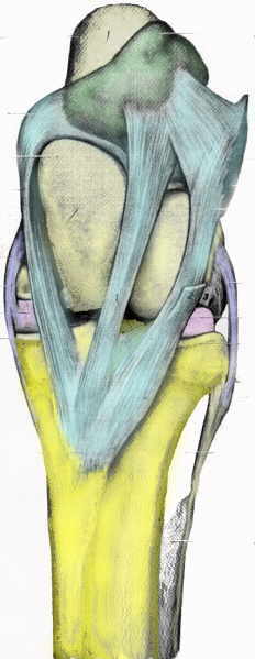
Front View of Left Stifle
Anatomy and Function
The stifle is a joint that actually represents the articulation of three bones: the femur at the top, the tibia at the bottom, and the patella out front. This is the same joint as your knee, complete with a knee cap: the patella. This is a very complicated joint with three joint compartments and many ligaments. From the front the patellar ligaments dominate. Looking at the picture to the right you see three ligaments originating from the patella and inserting on the tibia. Not shown is that the top of the patella is connected to the large and powerful quadriceps muscle group responsible for holding up the horse and helping to advance the leg.
Stifle Lock
Of particular interest is the medial (inside) patellar ligament. It is the near ligament in the picture to the right. See how it is hooked up over the bony prominence on the femur? By catching the ligament at this point the horse can lock the hind leg in a weight bearing position. On some horses the leg becomes unintentionally locked and may require surgery to correct.
Meniscus
Because the stifle bears so much pressure, the femur and tibia are separated by two very thick ligaments, the menisci, which acts as a lubricated cushion.
Legend for images above and below:
Femur
:
pale yellow,
Tibia
:
bright yellow,
Patella
(front view only): green,
Patellar ligaments
(front view only): light blue,
Menisci
:
magenta,
Collateral ligaments
:
blue,
Cruciate and posterior ligaments
(rear view only): orange.
Rear View of Stifle
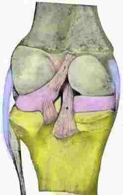
From behind the menisci are particularly visible. Also, seen are the insertions of the two cruciate ligaments. These ligaments span the femur and tibia, connecting the two together in a criss cross fashion. Attempts to advance the leg against resistance can result in rupture of these ligaments.
Localization and Diagnosis
Introduction
»
Anatomy
»
Localization and Diagnosis
»
Common Diseases of the Stifle
»
More Info & Discussions
To read more on this topic become a member of
Horseadvice.com! Your membership gets you instant access to this and over 600 equine articles on our site. Other benefits of your membership include participation in our discussion boards and access to our one button PubMed search tool for each topic.
Horseadvice.com educates you to be a more knowledgeable horse owner which leads to healthier horses and save you money, we guarantee it. Come Join Us!
 .
.