Distal Sesamoidean Ligaments of the Horse
by Robert N. Oglesby DVM
Introduction
Introduction
»
Functional Anatomy
»
Superficial (Straight) & Middle (Oblique) Sesamoidean Ligaments
»
Deep (Cruciate and Short) Sesamoidean Ligaments
»
More Info & Discussions
With improved diagnostic imaging techniques injury to the distal sesamoidean ligaments is becoming recognized as a more common cause of lameness in horses than was previously thought. Most often the oblique (middle) and/or straight (superficial) distal sesamoidean ligaments are involved. Desmitis (strain or sprain) of the deep and and short ligaments is a very rarely diagnosed condition. The location of the distal sesamoidean ligaments is deep to the flexor tendons which makes diagnosis from clinical exam alone difficult. Following localization of the lameness to the pastern it requires ultrasound or magnetic resonance imaging to visualize the damage and it's extent.
This article describes these ligaments and has images of the ligaments to give a better understanding of the anatomy of this area.
Functional Anatomy: part of the suspensory appartus
Introduction
»
Functional Anatomy
»
Superficial (Straight) & Middle (Oblique) Sesamoidean Ligaments
»
Deep (Cruciate and Short) Sesamoidean Ligaments
»
More Info & Discussions
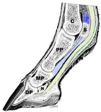
This is a view of the pastern and fetlock cut in half but just off the center, this is called parasagital.
Key to Image
- Cannon Bone: C
- Sesamoid Bone: S
- Proximal Phalanx: PP
- Middle Phalanx: MP
- Distal Phalanx: DP
- Navicular Bone: N
- Superficial flexor tendon: green
- Deep flexor tendon: yellow
- Proximal Suspensory ligament: blue
- Distal Sesmoidean Ligaments: light blue
- Superficial: Large outermost bundle with insertions on the MP
- Middle: The bundle in the middle and inserts in the middle of the PP.
- Deep: Short small ligaments that inser on the proximal aspect of the PP.
- Extensor tendons: magenta
Notice you can see a sesamoid bone which would not be seen if cut the pastern right down the middle. It is easy to appreciate here how the flexor tendons and the suspensory unit, consisting of the suspensory ligament, sesamoid bones, and distal sesmoidean ligaments help support the fetlock. Less well appreciated is how this unit, through the action of the distal sesmoidean ligaments, stabilize the fetlock and pastern joints. If you look carefully you can see there are 3 main groups of distal sesmoidean ligaments, colored light blue.
Superficial (Straight) & Middle (Oblique) Sesamoidean Ligaments
Introduction
»
Functional Anatomy
»
Superficial (Straight) & Middle (Oblique) Sesamoidean Ligaments
»
Deep (Cruciate and Short) Sesamoidean Ligaments
»
More Info & Discussions
To read more on this topic become a member of
Horseadvice.com! Your membership gets you instant access to this and over 600 equine articles on our site. Other benefits of your membership include participation in our discussion boards and access to our one button PubMed search tool for each topic.
Horseadvice.com educates you to be a more knowledgeable horse owner which leads to healthier horses and save you money, we guarantee it. Come Join Us!