The Collateral Cartilages and Sidebone in Horses
by Robert N. Oglesby DVM
Introduction
Introduction
»
Anatomy
»
Causes
»
Symptoms
»
Diagnosis
»
Treatment
»
Prognosis
»
More Info & Discussions
Each hoof has two collateral cartilages that lie to each side of the coffin (toe) bone of the horse. Through flexion and pressure changes, the CC's are likely ot help provide circulatory support to the foot particularly when in motion. A common condition of horses is to have the cartilages to calcify and enlarge. Because the upper edge lies above the hoof wall this can be seen and palpated and is called sidebone. This occurs most often in the front feet and may be related to conformation or trimming. Even when present it is not a common cause of lameness but there are exceptions. This article discusses the anatomy of the collateral cartilages, the identification of sidebone, and the diagnosis of lameness associated with sidebone. Also discussed is the treatment of sidebone related lameness and links to further information on this condition provided.
Anatomy
Introduction
»
Anatomy
»
Causes
»
Symptoms
»
Diagnosis
»
Treatment
»
Prognosis
»
More Info & Discussions
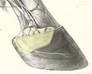
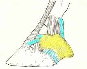
 yellow = collateral cartilage
|
 blue = ligaments attaching the collateral ligaments to the surrounding digital bones
|
The collateral cartilages lie beneath the skin, corium, and coronary venous plexus of the foot
(see yellow areas in image to the right). They are essentially L shaped with the horizontal bar projecting axially toward the middle of the foot. The upright part of the L is rhomboid- shaped with a convex surface facing the outside of the foot and a concave surface is facing toward the midline of the digital cushion. On the outside of the lateral cartilages are the extensive venous plexus, laminar dermis, and inner layer of the hoof wall. Several ligaments attach the collateral cartilages to the bones of the digit and the navicular bone
(see blue areas in image to the right).
The lateral cartilage is primarily hyaline cartilage, and in horses older than 4–5 yr, the medial boundary begins to develop fibrocartilage. On freshly sectioned specimens, the hyaline and fibrocartilage can easily be distinguished by the white and yellowish coloration of the respective cartilage. This development gradually creates a thickened lateral cartilage that can extend upward to more than 1.5 cm as a mean thickness along its length from the wings of the distal phalanx to the heels. Between the lateral cartilage and the DDFT, a robust fibrocartilaginous ligament becomes evident, which extends proximally to the level proximal to that of the navicular bone.
The morphological features of the lateral cartilages vary greatly from horse to horse in shapes and thickness, presence or absence of a fibrocartilaginous axial projection, and extent of the blood supply inside and outside of the cartilage. The variation of these morphological features appear to be mainly determined by environmental factors rather than by genetics. A robust ligament lying between the DDFT and the lateral cartilage is evident in the "good-footed" horse, whereas in other horses, only thin fibrous connections are evident between these two sites. Interestingly, in those feet in which the lateral cartilages begins to ossify in a palmar direction, this ligament may eventually become enveloped within the newly formed bone and may not be very functional.
Causes of Sidebone
Introduction
»
Anatomy
»
Causes
»
Symptoms
»
Diagnosis
»
Treatment
»
Prognosis
»
More Info & Discussions
To read more on this topic become a member of
Horseadvice.com! Your membership gets you instant access to this and over 600 equine articles on our site. Other benefits of your membership include participation in our discussion boards and access to our one button PubMed search tool for each topic.
Horseadvice.com educates you to be a more knowledgeable horse owner which leads to healthier horses and save you money, we guarantee it. Come Join Us!
 yellow = collateral cartilage
 blue = ligaments attaching the collateral ligaments to the surrounding digital bones