Developmental orthopedic disease (DOD) is a general term for any of the diseases of the musculoskeletal system that occurs during development and growth. This may occur in the fetus or the growing horse and may involve joints or other structures. Osteochondrosis (OC), a type of DOD, is a term for any of a group of diseases of the growth plates or ossification centers in the joints of young horses. Generally there are three forms of OC that are differentiated by their radiographic appearance. To try and make all these letters sensible, here it is in outline form:
-
Developmental Orthopedic Disease (DOD)
-
Angular deformities these are legs crooked when viewed from the front.
-
Flexural deformities are legs that appear contracted or weak when viewed from the side.
-
Physeal Dysplasia (Physitis) are legs that have swellings around the joints.
-
Ostechondrosis
-
Osteochondritis dissecans: defects in the cartilage surface.
-
Subchondral bone cysts: defects in the bone underneath the joint.
-
intraarticular bone fragments: also called joint mice, they are small pieces of bone or calcified cartilage in the joint space.
Osteochondrosis type diseases are all characterized by degeneration or necrosis of small parts of the epiphysis. This degeneration is sometimes followed by abnormal re-ossification.
All of the diseases under the term OC are defects that create weakness in the joint. When the weakness is such that exercise causes further trauma and damage lameness results. The symptoms of OC can be variable, with some cases causing no problems. Other cases can result in permanent lameness. OC is a lameness of young, growing horses. Usually the onset of lameness is subtle, becoming more obvious as the horse begins working. This article contains possible causes, diagnosis, treatment, prognosis, and has links to forum discussions and other resources on the Internet.
During growth bones elongate from the growth plates. These growth plates are usually just adjacent to the joint and joint surfaces made of cartilage. Growth plates represent a border between the cartilage on the end of the bone and the long calcified bone that makes up the shaft. Cells reproduce in the growth plate and continually differentiate. One side becomes more cartilage that lines the joints and the other becoming bone. It is the transission to bone that allows the bone to elongate. In young foals this is a very dynamic process of growth, modeling, remodeling, and even repair.
One critical event implicated as the primary event in OC lesions is the remodeling blood supply to the growth plate. As the bone enlongates the blood supply to the growth plate has to switch around from the end of the bone to the long portion of the bone. It appears that at this stage the blood supply to the growth plate occasionally fails to make a successful transition resulting in focal areas of arterial and bone necrosis. This may be the initiating event to many OC lesions. Why this failure occurs remains uncertain but there are some factors that would seem to contribute:
- Rapid or uneven growth
- Nutritional deficiences
- Trauma
- Genetic Influences
This is unlike the adult or even yearlings when these processes slow down remarkably and come to almost a standstill by maturity.
|
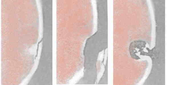
Examples of OC lesions.
The bone and cartilage have been cut to demonstrate the lesion.
From left to right: cartilage flap, cartilage fragment, cyst
|
In developmental diseases of the joint and cartilage, the cartilage of the joint surface or the adjacent bone has developed abnormally. There are debates over how it becomes abnormal or even what should be considered abnormal. One report suggests that all foals younger than 5 months may have some OCD lesions that for the most part resolve over the next 5 months. The question may be why do some foals not heal?
Trauma & Lack of Exercise
There is no doubt that both acute and chronic trauma are important initiating cause of clinical OC. But the relation between trauma and OC is complicated. For instance exercise, which could be considered a form of trauma, is important for proper cartilage growth, modeling, and strengthening in the first year of life. Long term forced rest induces a permanent weakening of the cartilage predisposing to OC lesions. So proper development requires exercise (trauma) to strengthen the cartilage but too little, too much, or poorly designed exercise regimens may induce significant lesions that persist.
Genetics
The larger, heavier breeds are at increased risk for OC. Since the incidence is increased in certain lines and breeds there are genetic factors at work. Also to be considered is the increased trauma caused by heavier bodies. correlation is weak enough that there are no recommendations to not breed an animal on the basis of a previous foal with OCD.
Heaviest alone however is not the only genetic factor as we see different lines of same breed horses that have OC lesions predisposed in different joints. More and more it appears that history of OC in a breeding horses family should seriously be considered a reason not to breed.
The Candidate Gene XIRP2 at a Quantitative Gene Locus on Equine Chromosome 18 Associated with Osteochondrosis in Fetlock and Hock Joints of South German Coldblood Horses
J Hered. 2009 Mar 20.
Wittwer C, Hamann H, Distl O.
the Institute for Animal Breeding and Genetics, University of Veterinary Medicine Hannover, Foundation Bünteweg 17p, 30559 Hannover, Germany.
A whole-genome scan for radiological signs of osteochondrosis (OC) and osteochondrosis dissecans (OCD) in South German Coldblood (SGC) horses using 250 microsatellite markers identified a genome-wide significant quantitative trait locus (QTL) for fetlock OCD and a chromosome-wide QTL for hock OC on Equus caballus chromosome (ECA) 18 at a relative position of 45.9-78.2 cM. The aim of this study was to analyze associations of single-nucleotide polymorphisms (SNPs) in candidate genes for OC in this QTL region using 96 SGC horses. The OC-QTL on ECA18 could be confirmed and narrowed down to an interval of 13 Mb between GALNT13 and Xin actin-binding repeat containing 2 (XIRP2). SNPs in the XIRP2 gene were significantly associated with fetlock OC, fetlock OCD, and hock OC. The significant associations of SNPs in XIRP2 could be confirmed in linear animal models controlling for systematic environmental and residual quantitative genetic effects. The significant additive genetic effects of the intronic SNPs (AJ885515:g.159A>G, AJ885515:g.445T>C) in XIRP2 were 0.15 (P = 0.01) for fetlock OC, 0.27 (P = 0.01) for fetlock OCD, and 0.15-0.16 (P = 0.01-0.02) for hock OC. Homozygous (A/A or T/T) and heterozygous horses were at a 1.3- to 2.4-fold higher risk for fetlock and hock OC. These results suggest that dominant variants of XIRP2 may be involved in pathogenesis of equine OC.
|
Nutrition
Because of frequently published articles and advertisement focused on nutritional research and the selling of products, many horse owners think that OC is primarily a disease due to nutritional deficiencies and this is simply not true. Though there are many hypotheses of nutritional causes of OC, there is little clear proof for most of them. Nowadays nutritional deficiency is not that common other than perhaps over nutrition. And here there is evidence that rapidly growing foals are more prone to OC lesions. It remains uncertain whether this is a nutritional or genetic problem and most likely both. Besides the increased stress of heavier foals there has been the idea that rapid growth may not allow for proper healing of an OC lesion resulting in a weak spot in the cartilage.
Other nutritional factors that have been associated with OC:
-
Experimentally OCD can be induced with decreased copper intake or excessive levels of zinc in the diet.
-
There have been correlations made of an increased incidence on farms with uneven levels of calcium and high phosphorous levels.
-
Uneven nutrition, like that associated with turnout on rapidly growing lush pasture may contribute to problems.
Other Causes
Though rare there are incidences of severe, systemic, multifocal OC cases in horses and have been associated with:
History of a OC Lesion
With a lot of "perhaps and maybes" here is a possible scenario of how an OC lesion might form. A foal of a large breed is born with small defects in the cartilage of a long bone. He may have a few more than many of his breed but normally these repair over the first year of life. The foal, receiving heavy nutrition and growing rapidly is stalled 12 hours daily, during which time the cartilage does not receive proper stimulus so is not as strong as some of his cousins who have nearly 24 hour a day turn out. On top of that being up for 12 hours most days leads to a roaring hard romp around the pasture when first turned out. During one of these episodes the foal bruises the cartilage.
This bruising leads to abnormal cartilage growth. This area thickens but still is a bit weaker than the surrounding cartilage. There are no blood vessels in the cartilage and it receives nutrition by diffusion from the joint fluid. If the cartilage becomes too thick, it cannot receive adequate nutrition by this mechanism and continues to weaken further. In time the area develops fissures, and the fissure become a flap, then the flap breaks off, leaving a defect in the cartilage surface. By this time the foal has become a young adult. During the past year the owner has noticed the horse occasionally just a little off but often fine by the next morning. Light training is begun and by the second week a persistent lameness that worsens during work develops. Unfortunately a frequent sequelae to clinical OC is arthritis and eventually degenerative joint disease. A schedule for a endoscopic examination of the joint the pain refers to is scheduled for next week.
 .
.